Helping aspiring clinicians understand a virtual heart before they work with a real one
Jonathan Awori, MD, MS, MFA, isn’t embarrassed to say it took him a long time to completely understand the intricate workings of the heart. He says he isn’t alone; many aspiring doctors and nurses don’t immediately grasp the heart’s complexity from two-dimensional textbooks or even 3D-printed models.
Now that he’s a pediatric cardiology fellow at Boston Children’s, Awori wants to make learning the heart anatomy easier and more of a lasting experience. He has a strong hunch, one he wants to confirm with a study, that virtual reality (VR) can do what no textbook can: introduce the heart almost as it really is.
“People enjoy virtual reality. It’s a cool tool and fun,” he says. “But what we’re trying to find out is if it actually helps enhance learning, if it gives you a real sense of what the heart does.”
Rotating and slicing the virtual heart
For years, medical trainees relied on words, photos, illustrations, and physical models to learn the structure and functions of the heart. They still lean on those mediums, but technology now has a key role in instruction. Trainees closely study MRIs and CT scans not only to understand the heart but also to prepare for when, as licensed practitioners, they will use those images to diagnose patients and create treatment plans.
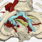
They might even someday work for a heart program that pushes the boundaries of digital technology to diagnose and treat patients. Boston Children’s, for example, uses 3D modeling software to capture unique perspectives of a patient’s heart scans and create individualized surgery plans for complex conditions.
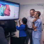
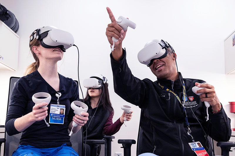
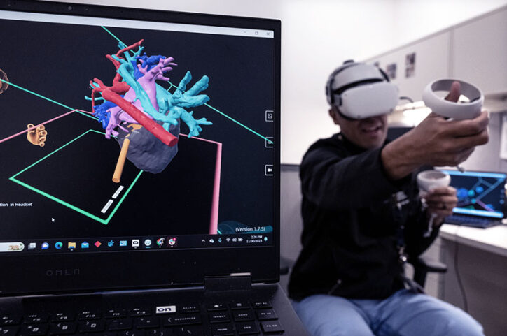
Like 3D modeling software, VR offers distinctive views of the heart. It makes the heart, Awori says, look realistic with a “high degree of detail, especially the heart chambers where you have vessels coming and going.” The virtual heart can be rotated to reveal a variety of angles, and a user can slice through it, take it apart, and make sections disappear to get a closer view of hard-to-see components.
“I can make the front of the heart invisible so I can see the back. Detailed effects like that make the technology valuable and I think pretty effective,” he says. “One of the things we know about learning theory is adults like to manipulate and have agency over their learning space.”
Going beyond fun to see if VR helps learning
Awori enjoys seeing trainees have fun with VR because it makes them feel at ease, but that’s not enough to gauge whether it’s an effective learning tool. To determine that, he’s about to embark on his second study of VR in heart education in the past few years.
His first study, conducted while serving as a chief resident at Seattle Children’s Hospital, found that residents and nurse practitioners preferred VR views of the heart over 3D-printed models to understand the heart’s anatomy and how it works.
Now part of Boston Children’s Cardiology Fellowship Program, Awori is starting another study that will try to look beyond VR being an enjoyable learning experience by objectively assessing whether trainees actually learn more. With the help of Jeffrey Jacobson and Angelina Gu from Boston Children’s Immersive Design Systems laboratory, Awori hopes that at least 50 Boston Children’s cardiology fellows, nurse practitioners, and physician assistants can participate in the study, which comes in three steps:
- an initial test of their heart knowledge
- followed by a 30-minute session of exploring the heart with VR
- another test with subjective questions about the VR experience alongside objective questions about heart anatomy and functions
Introducing a new visual theater to heart trainees
If VR is accepted as an educational tool to understand the heart, it could drastically reduce the time it takes trainees to learn, Awori says. For instance, fully understanding how an echocardiogram presents viewpoints of the heart requires months of repeatedly looking at those images.
But VR doesn’t have to be the only option for learning, he says. “Maybe it’s not practical to use that every time they take care of a patient, but if we can find different iterations of virtual reality, that kind of representation can also help learners.”

Awori has a master’s degree in theater and professionally acted in many plays and musicals before entering medical school. He likes to point out that the Greek word for “theater” originated from a Greek verb that means “to view.” And that’s what he envisions with VR in cardiology: “How do you help people see differently?”
Learn more about the Department of Cardiology.
Related Posts :
-

From aerospace to the OR: 3D modeling improves surgical planning by revealing details of patients' hearts
One of the most important tools for complex heart surgeries at Boston Children’s isn’t even in the operating ...
-
Research aims to pinpoint genetic connection between autism and heart disease
Cardiology and neurodevelopmental researchers have more questions than answers about the possible genetic links between congenital heart disease (CHD)&...
-
Finding a way to help newborns who can't immediately have heart treatment
Newborns with complex congenital heart defects (CHD) and pulmonary overcirculation often need treatment as soon as possible. Unfortunately, ...
-
Reconstructing a chest wall, one virtual step at a time
It takes a village of clinicians and engineers to reconstruct a chest wall. It also takes a lot of 3D ...