A new lens on cardiac surgery could help prevent arrhythmia
Sometimes, a change in perspective can lead to a medical breakthrough.
A type of microscopy typically used to detect cancer and other diseases has been adapted to reveal the location of unseen conduction tissue around the heart. The Boston Children’s clinician behind this innovation now aims to prove the safety and effectiveness of fiber-optic confocal microscopy in cardiac surgery so that it can be an indispensable tool for diminishing the risk of conduction tissue injury that causes patients to rely on pacemakers.
“We want this technique to be a standard part of every heart operation,” says Aditya (AK) Kaza, MD, a cardiac surgeon in the Benderson Family Heart Center’s Department of Cardiac Surgery. “We want surgery to improve and not disrupt a child’s health.”
Finding a new purpose for powerful technology
Cardiac surgeons have to be cautious as they operate around the conduction system — a network of cells and electrical signals that control the beating of a heart. Conduction tissue is not visible to the naked eye, nor to imaging technologies in operating rooms, often prompting surgeons to say they rely on an “eye of faith” when working near it.
Conduction systems vary from person to person, but they’re especially difficult to pinpoint in patients with complex congenital heart defects (CHD). When surgeons can’t locate conduction tissue during surgery, they risk accidentally injuring it. This can cause heart block, a type of arrhythmia that often creates the need for a pacemaker and more operations.
Kaza has always wanted to solve the conduction system dilemma and saw an opportunity a decade ago when he was a cardiac surgeon at Primary Children’s Hospital at the University of Utah. Two academic colleagues at the university — biomedical engineering professors Robert Hitchcock and Frank Sachse — told Kaza their department had a new fiber-optic confocal microscope. “It became something of a laboratory curiosity as we figured out what to do with it,” recalls Hitchcock. “AK asked if it could be used to identify conduction tissue.”
A regular microscope shows only the surface of tissue. A confocal microscope offers a deeper, high-resolution view of tissue by blocking out-of-focus light and creating a singular source of light — a perspective that helps pinpoint endocrine and metabolic diseases at the cellular level. That kind of perspective, Kaza and his colleagues believed, could also be used to make distinctions of anatomy beneath the heart’s surface, pointing to the characteristics of a conduction system.
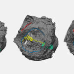
Differences in the appearance of cells point to location of conduction tissue
There are different types of confocal microscopes. The one used by Kaza’s team is the size and shape of a bendable straw, fitting ideally in a surgeon’s hands. Still, there was one big problem with bringing the technology into an operating room: It was linked to a large image-processing system. Most equipment in a surgical theater is portable and requires little space so that clinicians can move freely.
Kaza and the professors ultimately partnered with a medical device manufacturer to create a setup that won’t get in the way of cardiac surgeons. The handheld microscope connects to a six-foot-long cable stuffed with 30,000 fiber-optic strands that transmit high-powered visuals to an image-processing machine the size of a desktop computer.
The microscope looks about 50 millionths of a meter below the heart’s surface, or about half the thickness of a standard sheet of notebook paper. The microscope doesn’t reveal conduction tissue itself. Rather, it contrasts the visual differences of heart muscle cells. Regions of cells that are striated (long, thin lines) do not have conduction tissue, while cell areas that are reticulated (arranged like a net) are part of the conduction system. The idea is that when surgeons look at the different presentations of heart muscle cells, they will recognize that “messy” reticulated areas are spots to avoid.
Confocal microscopy in cardiac surgery trials shows ‘encouraging’ potential
Over the past decade, Hitchcock and Sachse worked closely with Kaza — even when he came to Boston Children’s — as he led studies of confocal microscopy’s effectiveness and safety in cardiac surgery. “We were initially concerned about how difficult it would be to steer the microscope through the human heart, but it’s actually pretty flexible,” Kaza says. Imaging takes two to five minutes after the heart is stopped and supported by a bypass machine. Surgeons then mark areas of conduction tissue with a marker so they can avoid accidentally injuring it.
The scope of their studies has broadened through the years: first on six sheep, then on six human patients, from infancy to age 21, who needed atrial septal defect repair. That study showed conduction tissue in the atrioventricular node and sinus node regions. But because the location of conduction tissue varies greatly depending on the type of CHD, Kaza and his team continue clinical trials. He and eight other Boston Children’s cardiac surgeons recently finished a “very encouraging” trial of 27 pediatric patients that revealed substantial differences in where conduction tissue is located in hearts with ventricular septal defects, tetralogy of Fallot, and atrioventricular canal defects.
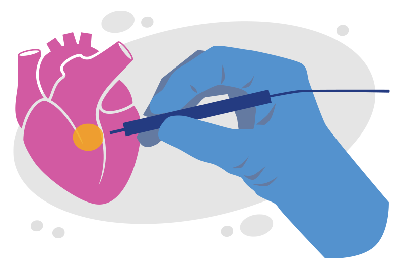
The confocal microscopy project isn’t the only Boston Children’s initiative trying to prevent surgery-induced arrhythmia. Benderson Family Heart Center cardiac surgeons, cardiologists, and engineers are testing the use of a catheter to find conduction tissue in hearts beating naturally during surgery.
The research involving Kaza’s team has taken years, but he believes the work will offer great rewards. “If you look at the treatment of cancer, it’s about being localized and precise to spare healthy tissue,” he says. “This approach to cardiac surgery is no different. Confocal microscopy can help surgeons be precise and locate conduction tissue so we can protect it.”
Learn more about the Department of Cardiac Surgery or refer a patient to the Benderson Family Heart Center.
Related Posts :
-

"Seeing" the unseen: A way to pinpoint elusive cardiac conduction tissue
When patients with congenital heart issues have an operation, surgeons have to proceed with an “eye of faith” as they ...
-
Two new approaches to identifying conduction tissue
Conduction cells in the heart are responsible for initiating contraction of the heart muscle. The inability to properly identify the ...
-
From South Africa to Boston: Kyleigh's four-year search for good heart health
Kyleigh Kista had three open-heart surgeries in just the first 18 months of her life. But instead of progressing, her ...
-
Wanting to give back, four heart patients now work as heart pros
They belong to a unique group. As children, they needed surgery to repair congenital heart conditions. Rather than being ...