Mending injured hearts: Lessons from newborns?
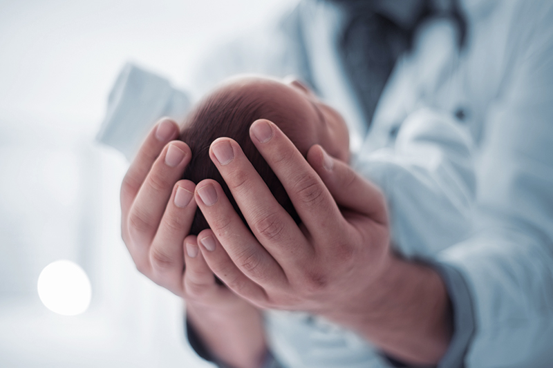
When the heart is injured, as in a myocardial infarction, the damaged heart muscle cannot regenerate — instead, scar tissue forms. Cardiomyocytes, the heart muscle cells that generate contractile force, are lost for good. Yet, in mouse models, the hearts of newborns regenerate readily after injury.
How are newborn hearts able to recover? What are the necessary ingredients for regeneration? Miao Cui, PhD, began asking these questions as a postdoctoral fellow in the lab of Eric Olson at the University of Texas Southwestern Medical Center. Continuing the investigations in her own lab in the Department of Cardiology at Boston Children’s Hospital, Cui has been finding factors that support heart regeneration. They include a pathway that cancers have co-opted to escape attack.
Could interventions reactivate some of these factors to treat cardiomyopathies in children and heart disease in adults?
An audit of the newborn heart
Early on, Cui and Olson identified a unique subpopulation of cardiomyocytes that retain regenerative capacity. Looking at RNA sequencing data from these so-called CM4 cells in a mouse model, Cui has identified several regeneration-friendly factors.
One, a transcription factor known as NFYa, is involved in mitochondrial metabolism and is key in activating genes involved with cell proliferation in the growing fetal heart. As recently described in Developmental Cell, loss of NFYa led to a decrease in immature, regenerating cells; an increase in mature, non-regenerating cardiomyocytes; impaired mitochondrial metabolism; and failure of the heart to grow.
Another transcription factor, Nrf1, regulates a genetic program involved in adapting to cellular stress. As Cui and colleagues detailed in Nature Communications, loss of Nrf1 prevented newborn hearts from regenerating. Conversely, increasing expression of Nrf1 in adult mouse hearts protected them from ischemia/reperfusion injury — a promising lead Cui is investigating further.
Heart regeneration, immunosuppression, and cancer
The latest work, co-led by Stephanie Vargas Aguilar, PhD, at UT Southwestern and published in Nature Cardiovascular Research, identified a surprising factor that helps the newborn heart regenerate: the PD-1/PD-L1 pathway. This well-known pathway suppresses the immune system; cancers use it to avoid immune attack by the body’s T cells. In oncology, it’s the target of many checkpoint inhibitor drugs.
In the newborn heart, activation of the PD-1/PD-L1 pathway maintains an immunosuppressive environment that enables regeneration, the researchers found.
“We found systematic downregulation of PD-1 and PD-L1 in the immune system after birth,” notes Cui, who also holds an appointment in the Department of Genetics at Harvard Medical School. “It is possible that, during the pregnancy, high PD-1/PD-L1 activity is beneficial for escaping the maternal immune response. However, once the mice are born, it is critical to quickly reduce the levels of PD-1/PD-L1 to prevent infections.”
When the researchers suppressed the pathway in newborn mice with a cardiac injury, they saw increased inflammation and T-cell activation and impaired heart regeneration.
“We surmised that activation of T cells is promoting the death of cardiomyocytes,” Cui says. She thinks the findings could also explain why cancer patients receiving immune checkpoint inhibitors often develop myocarditis.
Exploring therapeutic approaches
Cui’s next step is to investigate ways to up-regulate the PD-1/PD-L1 pathway in the heart through gene therapy or cell therapy (transferring newborn-like T cells) to get heart tissue to regenerate after a heart attack.
That’s just one potential therapeutic target Cui hopes to explore. Her lab aims to uncover additional factors through a systematic survey of genetic networks that regulate regeneration. The goal would then be to over-express them through gene therapy in animal models to see the effects on cardiac development and recovery from stress and injury.
Cui also hopes to apply her studies to human heart tissue in collaboration with Sarah Morton, MD, PhD, in the Division of Newborn Medicine. Morton’s research focuses on the genetics of congenital heart disease (CHD). The two plan to investigate whether patients with CHD have mutations in the genes Cui has connected with heart regeneration.
“I am looking forward to this collaboration,” says Morton. “Appreciating how genetic variation could contribute to cardiac regeneration and resiliency could help us understand differences in outcomes among patients with CHD and might enable new therapeutic options to improve their health.”
Learn more about Cardiology Research and the Neonatal Genomics Program.
Related Posts :
-

A new view of heart health: Mutations accumulate in the heart starting in childhood
Why do so many people get heart disease when they get older? We know that factors like high blood pressure ...
-
The people and advancements behind 75 years of Boston Children's Cardiology
Boston Children’s Department of Cardiology has more than 100 pediatric and adult cardiologists, over 40 clinical fellows learning the ...
-
In the genetics of congenital heart disease, noncoding DNA fills in some blanks
Researchers have been chipping away at the genetic causes of congenital heart disease (CHD) for a couple of decades. About 45 ...
-
Helping aspiring clinicians understand a virtual heart before they work with a real one
Jonathan Awori, MD, MS, MFA, isn’t embarrassed to say it took him a long time to completely understand the ...