Breakthrough study finds possible causes of cardiac fibrosis in HLHS

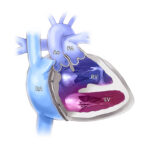
Many children who have hypoplastic left heart syndrome (HLHS) — one of the most severe congenital heart defects (CHDs) that affect circulation — also develop a condition in which the inner lining of their left ventricle thickens and further disrupts heart function.
The possible causes of the condition, a cardiac fibrosis known as endocardial fibroelastosis (EFE), have long been unknown, but new Boston Children’s research could finally bring clarity. Heart researchers discovered that endothelial cells, the cells that line the inner surface of blood vessels, are profoundly altered in EFE. The cells take on characteristics that stimulate scar formation and tissue remodeling. But that wasn’t all they observed.
“We also believe we found some important disease pathways that might be helpful to develop a new therapy, which could be helpful to HLHS patients,” says Department of Cardiology researcher Lili Zhang, MD, PhD, the principal investigator of the study. (Yang Yu, PhD, is first author; Kaifu Chen, PhD, and William Pu, MD, are co-corresponding authors).
Identity-shifting endothelial cells point to fibrosis
Zhang and Boston Children’s heart specialists have wanted to better understand the biological mechanisms driving EFE. Patients who have surgery to treat EFE experience different outcomes, so a clear cause could improve treatment.
Research outside of Boston Children’s had pointed to fibroblasts, common cells in connective tissue. Those findings made sense because the studied tissue was fibrous and had a limited number of blood vessels, Zhang says. But that research relied on technologies other than single-cell-level sequencing, an advanced method of identifying the variation between cellular signals and gene expressions that often show a cause.
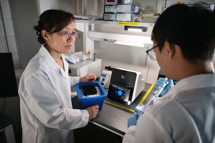
To identify cellular compositions and isolate abnormal signals that could potentially trigger EFE, the Zhang Lab performed paired single-cell RNA/ATAC sequencing on the heart tissue of three patients with EFE and tissue from three age-matched patients with healthy hearts. (Spatial transcriptomics was performed on two other EFE samples and three healthy tissue samples.) The findings — published recently in Circulation — surprised Zhang and team.
“We didn’t see an overabundance of fibroblasts,” Zhang says. “The big difference was the endothelial cells.” Endothelial cells were more abundant in EFE tissue than in healthy tissue.
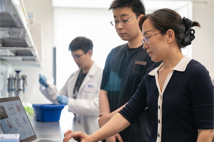
Zhang’s lab made another discovery. Separate groups of endothelial cells in EFE tissue seemed to have lost their true identity but at the same time hadn’t, Yu says. One group took on the identity of muscle-like cells and appeared to disrupt blood vessel growth. The other group had fibroblast-like features that could promote tissue scarring. Computational analysis by Yu showed the cells were actually in a transient state. Yu, Zhang, and their colleagues were onto something; next, they needed to learn what altered the cells’ gene expressions.
Finding pathways and hope for EFE treatment
The researchers went on to identify three dysregulated signaling pathways between endothelial cells and cardiomyocytes (the cells responsible for the heart’s beating). As a test, they then exposed healthy endothelial cells to abnormal signal molecules that are released by stressed cardiomyocytes in a cell culture environment. The stimulation caused the healthy cells to behave like the identity-shifting cells the team had discovered. Those once-healthy cells, Yu says, produced more collagen than normal, indicating the beginnings of the tissue scarring seen in EFE.
Because the lab worked with a small sample of tissue cells, they hope to soon study endothelial cells on a larger scale to advance their findings. More needs to be learned about the biological and mechanical connection between HLHS and EFE. Yet, even as a first step, Zhang says, the research shows promise that children with HLHS could someday receive effective targeted EFE treatment.
“We also hope to expand this to other diseases with fibrosis, not only congenital heart disease,” she says.
Learn more about the Zhang Lab and our approach to caring for children with HLHS.
Related Posts :
-

Biventricular repair linked to better outcomes in superior-inferior ventricles treatment
The complexity of superior-inferior ventricles — a condition in which one ventricle is positioned on top of the other — has often ...
-
First-of-its-kind pressurization test could improve Ross procedure outcomes
The Ross procedure is a preferred surgery to treat severe aortic valve disease. The procedure replaces the failing valve ...
-
Treating MAPCAs with unifocalization surgery and cardiology care
Children born with a rare form of tetralogy of Fallot (ToF) face a challenging type of congenital heart ...
-
Past patient outcomes could help single-ventricle surgery decisions
When considering whether a child who has a single-ventricle heart defect would benefit more from biventricular repair&...