Synapse ‘protection’ signal found; helps to refine brain circuits

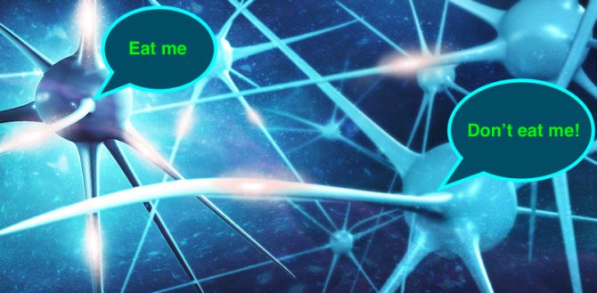
The developing brain is constantly forming new connections, or synapses, between nerve cells. Many connections are eventually lost, while others are strengthened. In 2012, Beth Stevens, PhD and her lab at Boston Children’s Hospital showed that microglia, immune cells that live in the brain, prune back unwanted synapses by engulfing or “eating” them. They also identified a set of “eat me” signals required to promote this process: complement proteins, best known for helping the immune system combat infection.
In new work published today in Neuron, Stevens and colleagues reveal the flip side: a “don’t eat me” signal that prevents microglia from pruning useful connections away. The signal, a protein called CD47, communicates with a receptor on microglia called SIRP alpha.
“We think this is the first evidence of a protective cue that microglia can read out that tells them not to prune,” says Stevens. “Our findings demonstrate that synaptic protection is necessary to ensure normal brain development.”
The neuro-immune connection
Like complement, CD47 also plays a role in the immune system, where it is part of a group of “don’t eat me” signals that prevent damage or removal of healthy cells. (In fact, some cancer immunotherapies work by inhibiting CD47, encouraging the immune system to attack cancer cells.)
“We asked, are any of these molecules expressed in the brain?” says Stevens. “Sure enough, CD47 is expressed very highly and is found throughout the brain. We think that, as seen following an immune challenge, the brain is using it as a protective cue, telling microglia not to prune a specific synapse. The brain and the immune system are sharing signals in a way that we’re only beginning to appreciate.”
CD47: the ‘yin’ to complement’s ‘yang’?
To model synapse development and pruning in the brain, Stevens and colleagues tapped a time-tested model of synapse refinement: the developing visual system.
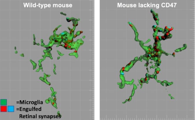
They found that during the time when synapse pruning in the visual system is at its peak, CD47 is enriched in the developing visual thalamus and localized to synapses. They also found that mice lacking CD47 (through a genetic deletion) had increased pruning activity by microglia and fewer synapses than normal mice, having lost the “don’t eat me” signal.

The findings add fuel to the idea that the brain has a balance of opposing factors that help fine-tune its connections — a yin/yang of sorts.
“The study is exciting because it suggests a possible cooperative interaction between ‘eat me’ and ‘don’t eat me’ signals that instruct microglia what to do when they see a synapse,” says Stevens. “As we start to delve deeper and identify new molecules and mechanisms by which microglia are pruning, it’s important to think how all these things fit together. It’s not one pathway, but a coordinated effort.”
A molecule to regulate activity-dependent eating
Another important aspect of the work is the link between CD47 and the activity of nerve cells, or neurons. Pioneering work over many decades has shown that the activity of neurons regulates the pruning process. But much less is known about how this happens on a molecular level.
Previous work in the Stevens lab showed that microglia, when given the choice, preferentially eat synapses from less active neurons compared to more active neurons. However, how microglia can tell these synapses apart remained unknown. The new study finds that in response to changes in neuronal activity, CD47 localization changes — with CD47 preferentially localized to synapses from the more active neurons. In the absence of CD47, microglia appear unable to distinguish different activity levels, as they no longer prefer to eat synapses from less active neurons.
“We think this is the first example of a molecule regulated by neuronal activity that can put the brakes on microglial engulfment,” says Stevens. “Exploring the mechanisms underlying this and determining whether it applies more generally to synaptic refinement in other brain regions will be an important aspect of future study.”
Preventing synapse pruning?
Though it’s still too soon to say, the study could have also implications for understanding and treating brain disorders. A number of neurodegenerative diseases such as Alzheimer’s disease and schizophrenia involve synapse loss, possibly through aberrant activation of pruning.
“Whether we can leverage this protective ‘don’t eat me’ signal to curb pathological synapse loss in disease is an open question,” says Daniel Wilton, PhD, second author on the Neuron paper.
Emily Lehrman, now at Clark + Elbing LLP, is the paper’s first author. The work was supported by multiple grants from the National Institutes of Health (NINDS). See the paper for a full list of grants and authors.
Related Posts :
-

The hidden burden of solitude: How social withdrawal influences the adolescent brain
Adolescence is a period of social reorientation: a shift from a world centered on parents and family to one shaped ...
-

The journey to a treatment for hereditary spastic paraplegia
In 2016, Darius Ebrahimi-Fakhari, MD, PhD, then a neurology fellow at Boston Children’s Hospital, met two little girls with spasticity ...
-

The dopamine reset: Restoring what’s missing in AADC deficiency
In March 2023, a young girl came to Boston Children’s Hospital unable to hold up her head — one striking symptom ...
-

The thalamus: A potential therapeutic target for neurodevelopmental disorders
Years ago, as a neurology resident, Chinfei Chen, MD, PhD, cared for a 20-year-old woman who had experienced a very ...


