Tough yet flexible: Biologically inspired adhesive may improve fetal surgery
In children with spina bifida, the neural tube that forms the spinal cord and brain doesn’t close during early prenatal development. That leaves the nerves of the spinal cord exposed to potential damage from fetal movement and the surrounding amniotic fluid. While surgeons can repair spina bifida soon after birth, the ideal would be to cover the exposed area during gestation, to protect the developing spinal cord.
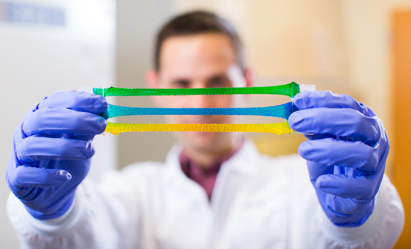
What’s been lacking is a surgical adhesive that itself can withstand fetal movement and growth and submersion in amniotic fluid, and is selective in what it sticks to. A new, nontoxic adhesive gel tested at Boston Children’s Hospital could fit the bill. Dario Fauza, MD, PhD, a general surgeon affiliated with Boston Children’s Maternal Fetal Care Center, believes it could improve prenatal surgery for spina bifida and perhaps other defects requiring a patch.
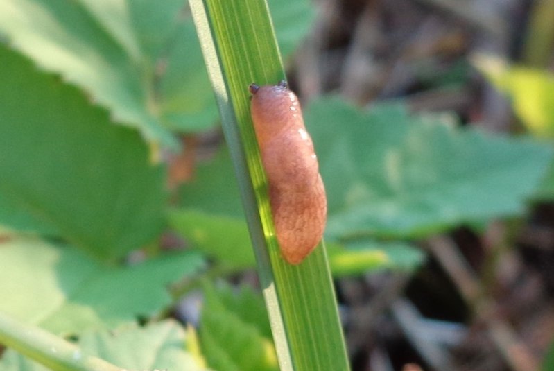
Inspired by the sticky secretions of the dusky arion slug, the gel was developed at Harvard’s Wyss Institute for Biologically Inspired Engineering and School of Engineering and Applied Sciences (SEAS).
“This collaboration was truly a team effort that brought together surgical and bioengineering expertise to address an important unmet clinical need,” said Benjamin Freedman, PhD, a postdoctoral fellow at the Wyss Institute and SEAS who co-created the technology. “We previously tested the tough adhesives in many living model systems, but none quite as complex as fetal surgery. We were blown away that the materials performed so well in this challenging setting.”
Putting a slug-inspired gel to the test
Learning of its good performance in sealing heart, skin, and liver tissues, Fauza decided to try it in fetal surgery, starting with spina bifida.
“The fetal environment, particularly in the setting of spina bifida, poses unique challenges for surgical adhesives,” says Fauza. “These include continued exposure to the chemically complex amniotic fluid and the fast growth rate of the fetus in the later stages of gestation.”
The new gel has two components: a hydrogel made of two bound polymer chains (alginate and polyacrylamide), linked to an adhesive polymer (chitosan). Boston Children’s surgical fellows Stefanie Lazow, MD, and Daniel Labuz, MD, tested the gel in the lab and in a prenatal rabbit model of spina bifida.
As reported last month in the Journal of Pediatric Surgery, the gel proved durable, strong, and flexible. Patches made from it adhered firmly around the edges of the spina bifida defect. As the fetus grew, the gel expanded, and remained in place in 70 percent of the animals tested. (Eventually, as the surgeons honed their technique, that rose to 100 percent.)
Avoiding tethered cord
With some conventional forms of surgical repair, the spinal cord can become tethered to the repair materials, potentially compromising spinal cord function. The new adhesive appears to reduce that risk.
“This new material allows for selective adhesion only to the periphery of the spina bifida defect, but not to the spinal cord,” Fauza says. “For adhesion to occur, both components must interact with the tissue, which can be controlled by the surgeon.”
The gel stood up to a battery of other tests, including submerging it in amniotic fluid and exposing it to hydraulic pressure mimicking the uterine environment. It kept wounds sealed even when wet, and even on moving surfaces. In the animal model, it survived the rigors of the intra-uterine environment, and the surgeons saw no tethering of the spinal cord.
We plan to explore this material’s use for other congenital anomalies exposed to the amniotic fluid, for example gastroschisis and tumors such as teratomas.”
– Dario Fauza
Finally, the adhesive appeared not to interact biochemically with fetal tissue, causing no inflammation or other toxicity. There was even some suggestion that the gel promoted wound healing, rather than simply acting as a patch.
Exploring noninvasive spina bifida repair
The team’s next steps will be to test the gel in larger animal models with longer gestation times (as opposed to 32-33 days in rabbits). They will also explore ways to introduce the gel less invasively, using a scope and a port site into the uterus, rather than open surgery.
“The hydrogel component can be rolled around an instrument, and the adhesive component is liquid, so both should be amenable to surgical placement via minimally invasive methods,” Fauza says. “We plan to explore this material’s use for other congenital anomalies exposed to the amniotic fluid, for example gastroschisis and tumors such as teratomas.”
Benjamin R. Freedman, PhD, and David J. Mooney, PhD, of the Wyss Institute and SEAS; Anna Rock, BS, of the Wyss Institute; and David Zurakowski, PhD, of Boston Children’s were coauthors on the paper. The study was funded by the Kevin and Kate McCarey Fund for Surgical Research and the National Institute on Aging of the NIH (F32AG057135).
Learn more about the Maternal Fetal Care Center.
Related Posts :
-

How the stars aligned for CeCe’s spinal cord tumor care
Last spring, 6-year-old CeCe was on vacation in Florida when she told her parents, Mike and Mary, that her neck ...
-
Thriving — not just surviving — as an adult with esophageal atresia
Willa Barnett has a story to tell — and it’s a big one. At 26, she’s experienced a slew of ...
-
Rh alloimmunization: A family’s experience across two pregnancies
After her second child was born 14 years ago, Erin was convinced she’d never have another baby. Not because of ...
-
How a meniscal transplant made me a Boston sports fan
I was in kindergarten when my knee started popping and cracking. My parents and I didn’t know it at ...