Laparoscopic approach to cloacal malformation repair found safe in eligible patients
A minimally invasive surgical approach called laparoscopic rectal mobilization and urogenital separation appears to be a safe alternative to open surgery in eligible patients with cloacal malformations. That’s the conclusion of a recent study by the team in the Colorectal and Pelvic Malformation Center at Boston Children’s Hospital.
A minimally invasive approach to urogenital separation
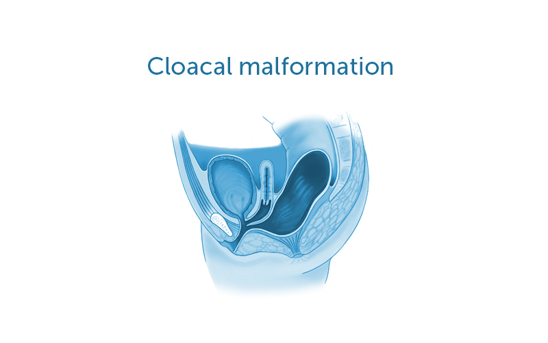
A cloacal malformation is a rare type of anorectal malformation that occurs when a female fetus’s rectum, vagina, and urologic structures form into one common channel. The choice of surgical repair strategy depends largely on the length of the baby’s common channel and urethra: Mid-sized to long common channel cloacas are challenging and often require laparotomy for dissection of pelvic structures. This large surgical incision allows surgeons to see all of the patient’s relevant anatomy, such as parts of the urinary tract, reproductive tract, and bowel. It also allows them to separate these structures from one another so that they can make individual openings.
Key takeaways
- Cloacal deformities are rare types of anorectal malformations that require surgical repair.
- The surgeons in Boston Children’s Colorectal and Pelvic Malformation Center have developed a laparoscopic approach to rectal mobilization and urogenital separation.
- This minimally invasive approach has the potential to result in faster recovery, shorter hospital stays, and less pain medication.
- A recent study by the team found this approach safe in patients with an average common channel length of 3.5 cm.
While laparoscopy has been used for rectal dissection, it had not previously been used for management of the urogenital structures. The center’s director, Belinda Dickie, MD, PhD, and associate director, Erin McNamara, MD, MPH, and their colleagues recently adopted a laparoscopic approach to rectal mobilization and urogenital separation.
This minimally invasive approach provides surgeons with a magnified view of the child’s anatomy. Because it requires smaller incisions, it also has the potential to result in faster recoveries, shorter hospital stays, and less pain medication.
Safe approach promises benefits for patients
To assess the safety of this approach, the team reviewed the records of nine children with cloacal deformities who underwent laparoscopic rectal mobilization and urogenital separation at Boston Children’s and elsewhere between 2016 and 2019. This included details on patient demographics, relevant anatomic lengths, length of operation, transfusion requirements, and perioperative complications.
The team found that the common channel length in these patients was 3.5 centimeters. There were no intraoperative complications, and transfusion requirements were minimal. The length of hospital stay was about six days. The findings were published in the November 17, 2020, issue of the Journal of Laparoendoscopic & Advanced Surgical Techniques.
“This minimally invasive approach allows us to visualize all structures in the abdomen and pelvis and separate them safely, offering the benefits of laparoscopic surgery,” says McNamara. “We will continue to follow these patients to see if there is improvement in other outcomes, such as continence.”
“Our surgeons are adept in both the open and laparoscopic reconstruction of cloacal anomalies,” adds Dickie. “We have adapted laparoscopy in certain patients as we feel it provides them with as good or better outcomes. Very few centers in the world have this breadth of experience.”
Learn more about the Colorectal and Pelvic Malformation Center.
Related Posts :
-

Answers for Aubree: Finding support for OEIS
Michelle and Stephen Strickland are used to having questions about their infant daughter, Aubree’s, health. After all, Aubree was ...
-
Innovative new shunt delivery system holds promise for treatment of fetal urinary tract obstruction
Urinary tract obstruction that occurs in utero can have serious consequences for the fetus. Such obstructions can block the flow ...
-
At the forefront of kidney stone removal: Innovative approaches transform patient care
No longer considered just an adult problem, kidney stones increasingly affect children as well. The majority of children who cannot ...
-
How the stars aligned for CeCe’s spinal cord tumor care
Last spring, 6-year-old CeCe was on vacation in Florida when she told her parents, Mike and Mary, that her neck ...