An ‘atlas’ of the choroid plexus across the lifespan
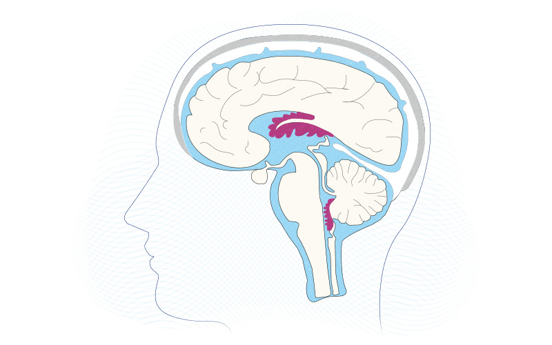
Once viewed merely as a producer of the cerebrospinal fluid (CSF) bathing the brain and spinal cord, the choroid plexus is now known to be a key player in brain development and immunity. These fronds of brain tissue, located in the CSF-filled brain cavities known as ventricles, secrete instructive cues into the CSF to regulate brain development. They also function as an important barrier between the brain and the rest of the body.
Key takeaways
Choroid plexus tissue, anchored in each of the brain’s ventricles and bathed in cerebrospinal fluid, is spatially inventoried with single-cell RNA sequencing, cataloguing cell types and gene expression patterns in each ventricle during early development, adulthood, and old age.
Maria Lehtinen, PhD, of Boston Children’s Hospital has done much of the pioneering work in understanding this once-obscure tissue. Her team recently showed, for example, that before birth, the choroid plexus can be a conduit for inflammation caused by maternal infection, paving the way for studies of neurodevelopmental conditions such as autism. Another study found that a protein in the choroid plexus helps reduce excess fluid levels in the brain, potentially providing a target for treating hydrocephalus.
In recent work published in Cell, Lehtinen, Neil Dani, PhD, and other colleagues at Boston Children’s and the Broad Institute created a cellular and spatial “atlas” of the choroid plexus during different life stages (early development, adulthood, old age). The map provides a benchmark to accelerate future studies investigating the lifelong regulation of this diminutive but influential brain structure.
“To fully understand the choroid plexus and its functions, we needed to identify its constituent cell types and their molecular composition,” says Dani, who was co-first author on the paper with Rebecca Herbst, a PhD student at the Broad Institute. “Such insights had been missing in the community.”
Extracting choroid plexus tissue
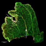
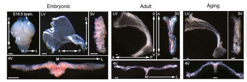
Isolating choroid plexus tissue from embryonic, adult, and aged mice was a job in itself. It required special micro-dissection approaches since these tissues lie deep within the brain’s ventricles.
“Figuring out how to dissect out intact third-ventricle choroid plexus was particularly challenging,” says Dani.
With tissue from each of the animals’ three ventricles in hand, the researchers performed RNA sequencing of more than 98,000 cells and cell nuclei to compare their gene expression profiles (what genes were turned on or off). This enabled them to catalog different cell types in choroid plexus from each ventricle, across different ages — the first time this has ever been done.
“We had previously done bulk RNA sequencing of choroid plexus tissue, but that was like putting everything in a blender,” says Lehtinen, who was co-senior author on the paper with Naomi Habib, PhD and Aviv Regev, PhD, at the Broad Institute. “We didn’t know which cells were secreting what. Sequencing cell by cell gives us a blueprint of what cells are where over the course of the lifespan. Knowing the different cell types, we can explore their roles and functions, how they talk to each other, and how the tissue is built.”
A choroid plexus inventory
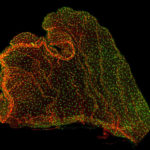
The investigations revealed a complex “architecture” of choroid plexus tissue specific to each ventricle. Gene expression varied especially among epithelial and fibroblast cells in the developing brain.
“We think the differences might have to do with helping guide the development of nearby brain regions,” says Lehtinen. “There are different ‘cocktails’ of factors secreted into different ventricles, in different patterns.”
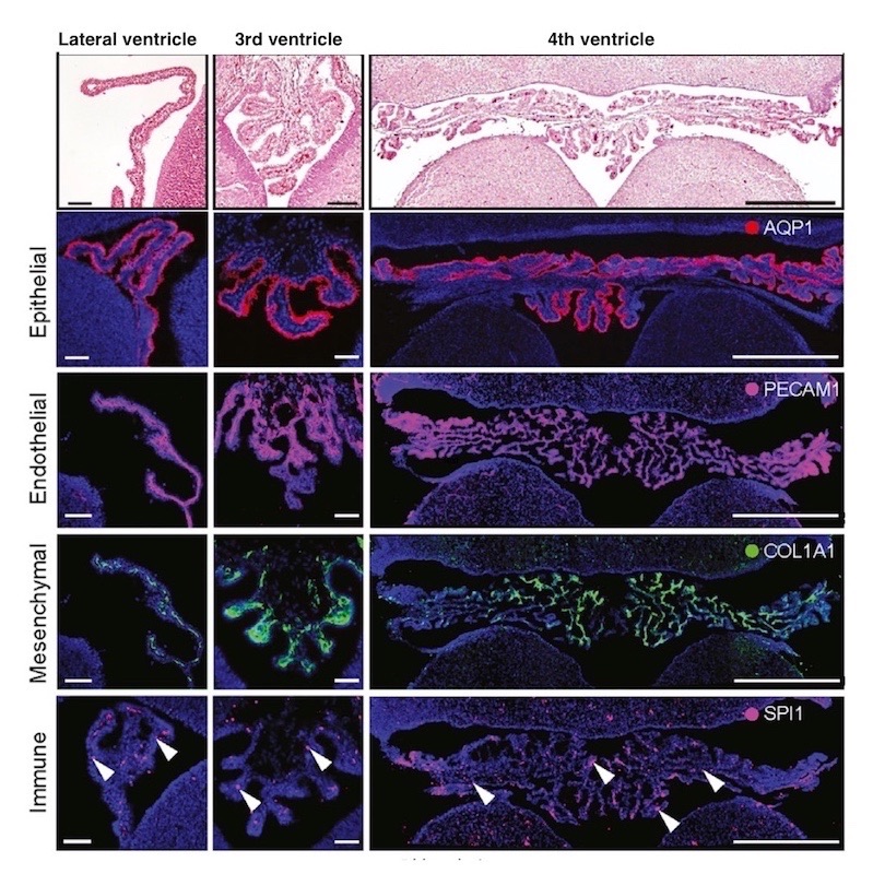
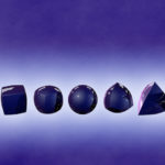
In addition to the core cell types shown above, the researchers found several neuronal subtypes. “Typically, the choroid plexus is not thought to have too many neurons,” says Lehtinen. “What they’re doing in the choroid plexus is still up for discussion and experimentation.”
The sequencing data also revealed differences in the cells’ secretions and molecular makeup.
“We were intrigued by the expression of insulin in the epithelial cells of the developing third-ventricle choroid plexus,” says Dani. “Whether this is a viable and functional central source of insulin in the brain needs further investigation. We also found that some epithelial and mesenchymal cells expressed ACE2 receptors, which the SARS-CoV-2 virus uses to enter cells. This underscores the need to understand how the choroid plexus functions not just in health but in disease.”
Defending the brain
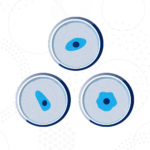
The team found a great deal of immune activity in the choroid plexus. Several kinds of immune cells reside there, most commonly macrophages. Immune signaling activity varied with age, with more inflammatory signals picked up in samples from aged brains.
“We can now start to analyze how different populations of immune cells are activated and how they respond to injury,” says Lehtinen. “This gives us a baseline and a set of molecular markers to start looking at.”
Other findings revealed the arrangement of arteries, veins, and capillaries in the choroid plexus, and their organization in relation to adjacent brain regions.
“Some of these vessels expressed blood-brain barrier proteins, which was not described before” notes Dani. “Once we mapped them, we found them to be continuous with arteries from adjacent brain regions.”
A therapeutic ‘window’?
With this resource in hand, the scientific community now has much to explore. Lehtinen and Dani believe that once the choroid plexus is better understood, it could provide a target for neurologic drugs.
“Gene therapy directed toward the CSF and choroid plexus could be an exciting therapeutic approach,” says Lehtinen. “It’s not going to fix everything, but it could have a sizeable impact.”
This work was initially funded by a Boston Children’s Hospital–Broad Institute collaboration grant. Other supporters include the National Institutes of Health, the Pediatric Hydrocephalus Foundation, the Reagan Sloane Shanley Scholarship, and the New York Stem Cell Foundation (see the paper for a full list).
Related Posts :
-

Exploring an unsung part of the brain: the choroid plexus
If you’ve never heard of the choroid plexus, you’re not alone. In fact, few neuroscientists know much about ...
-
The tiny choroid plexus protects the prenatal brain — but may also pass on inflammation from the mother
Floating in fluid deep in the brain are small, little-understood fronds of tissue. Two new studies reveal that these miniature ...
-
Single-cell sequencing reveals glioblastoma's shape-shifting nature
Glioblastoma, a cancer that arises in the brain’s supporting glial cells, is one of the worst diagnoses a child ...
-
Recommendations for reproducibility in stem cell research
The ability to program induced pluripotent stem cells (iPSCs) and drive their differentiation into a variety of neural cells is ...