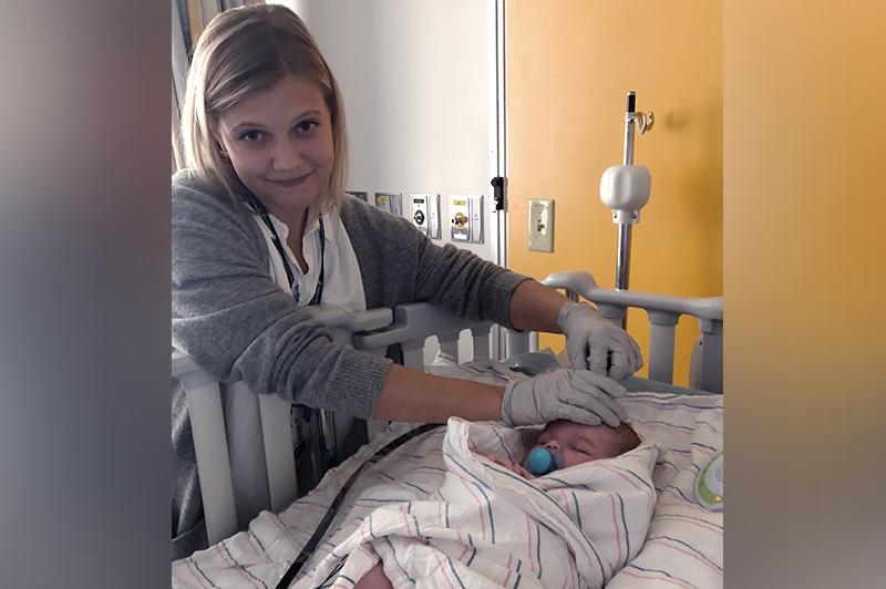
Bedside tech predicts newborns’ outcomes after therapeutic hypothermia

Hypoxic-ischemic encephalopathy (HIE), brain injury caused by oxygen deprivation around birth, is a common cause of admission to the neonatal intensive care unit. Therapeutic hypothermia is now becoming the standard treatment to minimize brain injury; cooling the newborn’s head or whole body for three days slows cellular metabolism and allows brain cells to avoid and recover from injury.
How do children fare after this treatment? A prospective multicenter study from Boston Children’s Hospital is the first to investigate the link between brain metabolism the day after hypothermia for HIE and children’s later neurodevelopment. It found that brain metabolism — specifically, the cerebral metabolic rate of oxygen consumption — is a sensitive predictor of babies’ cognitive and motor outcomes at 18 months.
Measuring newborns’ brain metabolism
Most current brain monitoring methods that use near-infrared spectroscopy (NIRS) look at oxygen saturation. But they don’t monitor oxygen consumption, which requires also measuring cerebral blood flow. Over the past two decades, Ellen Grant, MD, director of the Fetal-Neonatal Neuroimaging and Developmental Science Center, and her team at Boston Children’s have piloted a noninvasive bedside technology for measuring oxygen metabolism in newborns’ brains. It combines the more accurate frequency-domain form of NIRS (FDNIRS), which allows more consistent quantitative measurements from brain tissue, along with diffuse correlation spectroscopy (DCS), a new technology for measuring blood flow.
Together, FDNIRS and DCS quantify the cerebral metabolic rate of oxygen consumption (CMRO2) — an indicator of how well the brain is functioning.
“Oxygen consumption decreases during hypothermia, but when we return infants to normothermia, oxygen consumption reflects the number of healthy neurons and synapses, and is a better predictor of neurodevelopmental outcomes,” says Grant. “Other methods such as magnetic resonance imaging (MRI) can predict only the worst outcomes.”
Putting CMRO2 to the test
Led by Jason Sutin, PhD, and Pei-Yi “Ivy” Lin, PhD, Grant’s team prospectively enrolled 103 newborns admitted to Boston Children’s and two other Boston NICUs with suspected brain injury. Of these newborns, 58 received therapeutic hypothermia and were monitored and included in the analysis.
The team measured CMRO2 at the bedside during hypothermia, during rewarming, and after return to normothermia. They also scored the infants clinically and evaluated them with MRI and MR spectroscopy.
Oxygen consumption reflects the number of healthy neurons and synapses, and is a better predictor of neurodevelopmental outcomes.
CMRO2 was about 10 times more responsive to temperature changes during cooling than oxygen saturation from NIRS, indicating it is a more sensitive gauge of the effects of treatment. Among the 22 infants with 18-month follow-up data, CMRO2 after rewarming predicted cognitive and motor outcomes on the Bayley Scales of Infant and Toddler Development. In contrast, NIRS oxygen saturation or MRI showed no associations with outcomes.
CMRO2 was especially helpful in predicting neurodevelopmental outcomes of infants with mild HIE, who often had normal-looking MRI scans, says Grant.
The findings also suggest that mild HIE has a greater impact on children’s development than previously recognized. That could mean that hypothermia may benefit even newborns with mild HIE, a question the team wants to explore further. At the least, CMRO2 could help identify infants who would benefit from additional services.
From research to routine diagnostic
The National Institute of Child Health and Human Development has funded continued development of this technology. Grant, Lin, and Sutin are developing smaller, less expensive monitoring devices suited to clinical use. Clinicians in developing countries could also use such devices to assess cerebral oxygen metabolism in other conditions such as hydrocephalus.
“We want to take this from a research device and put into a form that could be used for routine diagnosis,” says Sutin. “This study is pivotal to prove that this more complex technology is clinically necessary.”
Learn more about the Fetal-Neonatal Neuroimaging and Developmental Science Center.
Related Posts :
-

Advancing mother-child health globally: Grace Chan MD, MPH, PhD
First in an ongoing series profiling researchers at Boston Children's Hospital. Globally, five million children die annually before the age ...
-

Could gene therapy relieve post-hemorrhagic hydrocephalus?
Premature infants, especially very low birthweight babies, are at risk for intraventricular hemorrhage. A frequent complication of these brain bleeds ...
-

Genetic sequencing may open doors for newborns with hypotonia
When a baby is born with low muscle tone (hypotonia), the future is hard to predict, and families have a ...
-

The sooner the better: An argument for fetal MRI before 16 weeks
To date, fetal magnetic resonance imaging (MRI) has been limited to the mid-second or the third trimester of pregnancy. This ...


