Surgical 3-D printing: 300 prints, 16 specialties and counting

3-D printing is rapidly becoming a part of surgical planning. Since July 2013, Boston Children’s Hospital’s 3-D printing service, part of the Simulator Program, has received about 200 requests from 16 departments around the hospital. It’s generated a total of about 300 prints, most of them replicating parts of the body to be operated on.
Most prints take between 4 and 28 hours to produce. The largest to date—an entire malformed rib cage—took 105 hours and 35 minutes to create and weighed 8.9 pounds. The smallest—a tiny tangle of blood vessels in the brain—took 4 hours and 21 minutes and weighed 1.34 ounces. Here is sampling of what’s been coming off the production line.
Note that production time includes segmenting the image (annotating the image data to indicate boundaries between anatomic structures), the actual printing, and post-processing (trimming, finishing and sometimes painting).
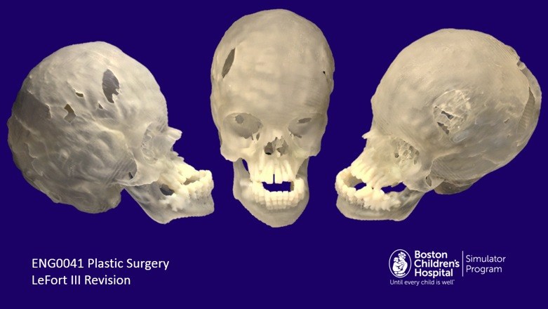
- Child’s age: 10 years
- Department: Plastic Surgery
- What was printed and why: This skull and mandible were printed to plan a LeFort III midface advancement, an operation that corrects certain facial anomalies by moving the upper jaw, cheeks, nose and orbital rims forward.
- Production time: Image segmentation, 6 hours; print time: 32 hours; post-processing, 1.5 hours.
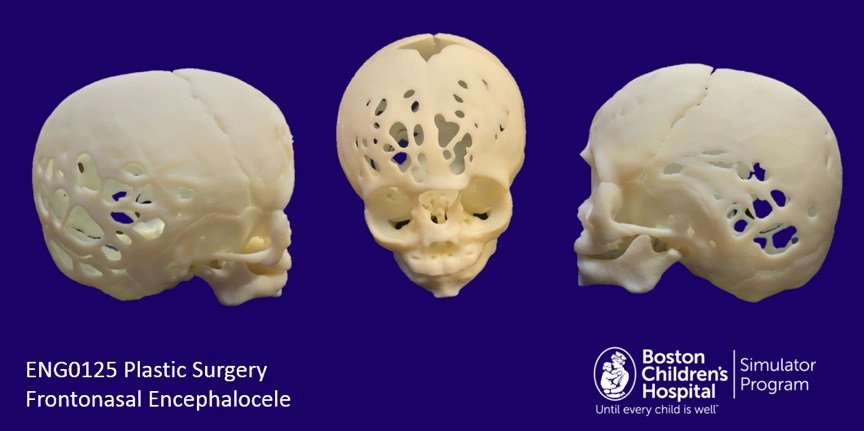
- Child’s age: 5 months
- Department: Plastic Surgery
- What was printed and why: This skull and mandible were printed to plan an operation to correct encephalocele, a condition in which abnormal openings allow brain tissue to protrude outside the skull.
- Production time: Segmentation, 2.5 hours; print time, 20 hours; post-processing, 1 hour
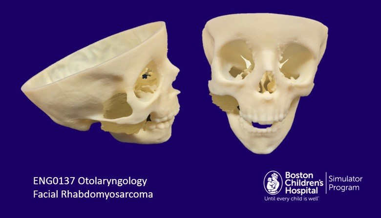
- Child’s age: 4 years
- Department: Otolaryngology
- What was printed and why: This child had a rhabdomyosarcoma, a cancerous tumor affecting soft tissues—in this case the jaw’s right masticator muscle. This pre-surgical print includes the rhabdomyosarcoma, in the right jaw, and the surrounding skull bone. It was made after 6 weeks of chemotherapy.
- Production time: Segmentation, 8.5 hours, printing, 22.5 hours, post-processing, 1 hour.
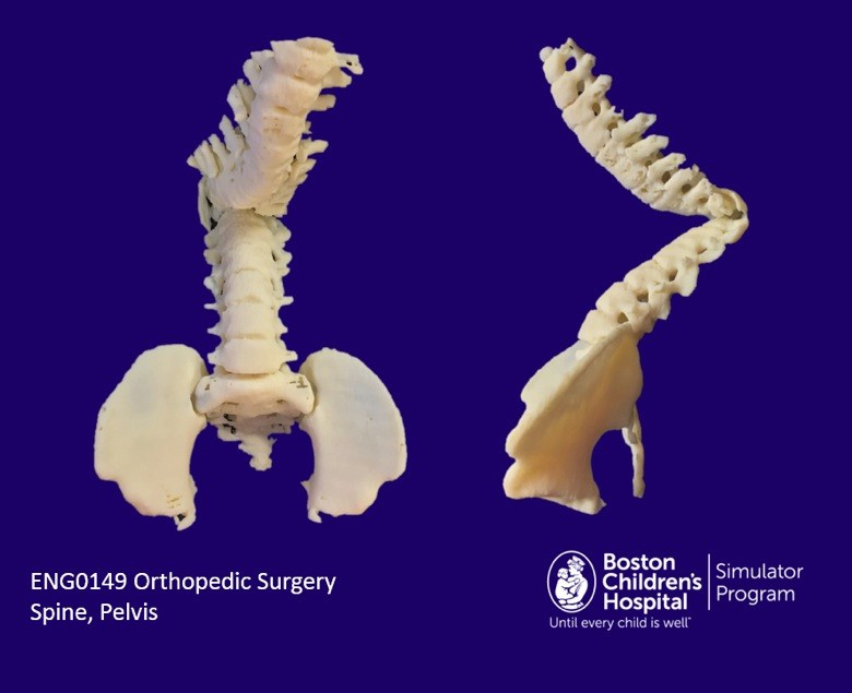
- Child’s age: 13 years
- Department: Orthopedic Surgery
- What was printed and why: This child has severe scoliosis (curvature of the spine) and myelomeningocele (the most severe form of spina bifida in which the backbone and spinal cord do not close during development). This replica of the thoracic spine, lumbar spine and pelvis was used to plan scoliosis surgery and to measure and bend rods prior to placing them surgically.
- Production time: Segmentation, 7 hours; printing, 45 hours, post-processing, 1.5 hours
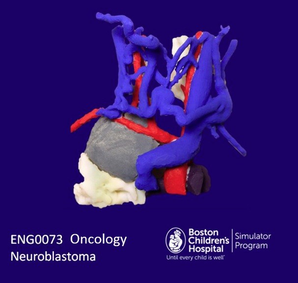
- Child’s age: 14 months
- Department: Oncology
- What was printed and why: This toddler was found to have a neuroblastoma—a large cancerous mass in his chest cavity around the spinal cord. It was compressing his trachea, making it difficult for him to breathe. This print shows the tumor, the connecting arteries and veins and part of the lung. It was used to help find a safe surgical approach to the tumor while avoiding the blood vessels.
- Production time: Segmentation, 13.5 hours; printing, 9 hours, post-processing, 2.5 hours
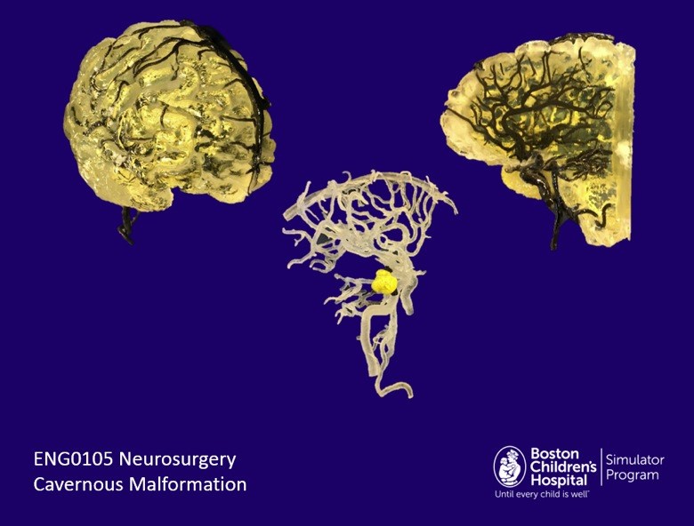
- Child’s age: 16 years
- What was printed and why: This teen had a cavernous malformation in his brain—a small, berry-like mass consisting of an abnormally thin-walled, expanded blood vessel. Untreated it could have caused seizures, weakness and other neurologic problems. The SIM program printed the malformation and surrounding vessels both alone (center) and embedded in the brain to help surgeons determine the best approach.
- Production time: Segmentation, 8 hours; printing, 20 hours; post-processing (malformation and vessels alone), 11 hours; post-processing (malformation, vessels and brain), 13 hours
The 3-D printing service is starting to be tapped for other applications—including rapid prototyping of innovative new devices to benefit kids.
Learn more about SIMPeds and SIMEngineering
Related Posts :
-

Thanks to Carter and his family, people are talking about spastic paraplegia
Nine-year-old Carter may be the most devoted — and popular — sports fan in his Connecticut town. “He loves all sports,” ...
-
AI-enabled medical devices are burgeoning, but many haven’t been tested in children
Medical devices that incorporate artificial intelligence and machine learning are proliferating. In 2013, the FDA approved fewer than 10 such devices; by 2023, ...
-
Going back to school with a chronic condition
Going back to school can be a time of excitement for many families: Your kids are looking forward to reconnecting ...
-
‘Zero place for mistakes’: Taking fetal surgery to the next level with simulation
The highly complex interventions involved in fetal surgery require exceptional skill, training, and experience. Beyond the procedures themselves, these surgical ...