The Beauty of the Brain
Every year, the Harvard Brain Science Initiative sponsors its Beauty of the Brain contest. This year, two Boston Children’s Hospital images are among the six winners drawn from a pool of forty submissions.
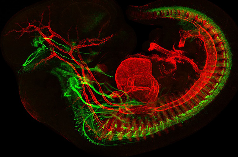
Above, Mary Whitman, MD, PhD, and Jess Bell, from the laboratory of Elizabeth Engle, MD, developed this image of a developing mouse embryo showing muscles in red and motor neurons in green. The Engle laboratory studies the genetics and molecular biology associated with eye disorders.
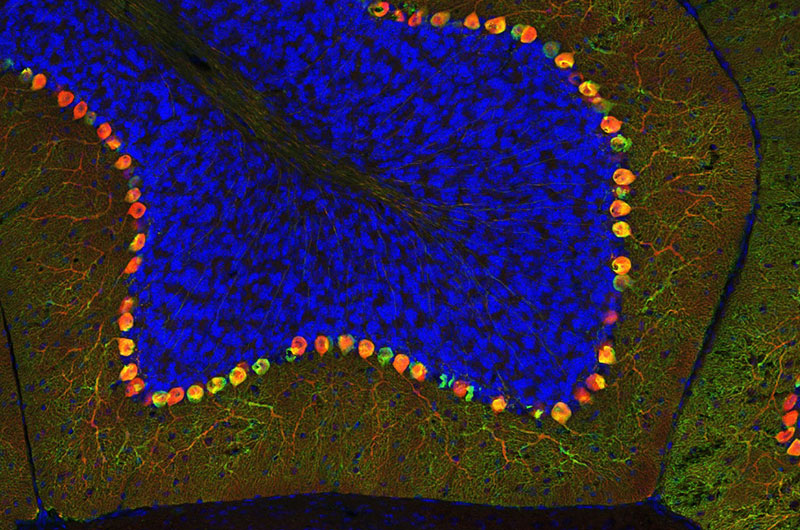
Below, Ellen DeGennaro, working in the laboratory of Christopher Walsh, MD, PhD, created this colorful image of a thin section of tissue from an adult mouse cerebellum. The image highlights blue nuclei, and Purkinje cells in red and green. The Walsh lab studies the genetics and molecular biology underlying human neurological diseases.
To view all the spectacular images visit the Beauty of the Brain
Related Posts :
-

Testosterone’s role in gut motility could open the door to targeting treatments for IBS and more
Irritable bowel syndrome (IBS) and other motility disorders can be challenging to treat, with current therapies focused mainly on easing ...
-
Hope in a new home: A family’s journey with HHT
When Yeiden Pérez Camacho imagines the future, he sees himself on a basketball court. At 13, he’s already an ...
-
Beyond average-based medicine: HIE as a blueprint for data-informed care
Historically, outcome prediction in medicine has followed a familiar formula: run a clinical trial, publish the results, guide care based ...
-
Promising advances in fetal therapy for vein of Galen malformation
In 2024, Megan Ingram* of California and her husband were preparing for the birth of their third child when a 34-week ...