The Beauty of the Brain
Every year, the Harvard Brain Science Initiative sponsors its Beauty of the Brain contest. This year, two Boston Children’s Hospital images are among the six winners drawn from a pool of forty submissions.
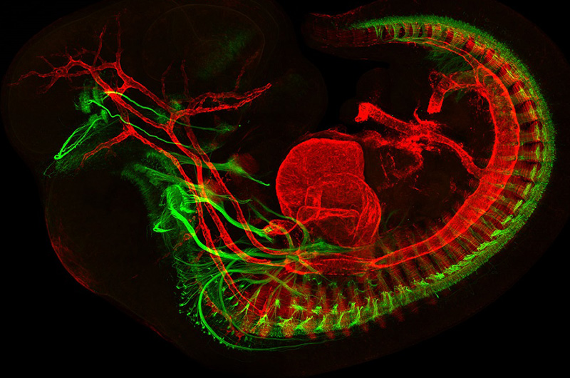
Above, Mary Whitman, MD, PhD, and Jess Bell, from the laboratory of Elizabeth Engle, MD, developed this image of a developing mouse embryo showing muscles in red and motor neurons in green. The Engle laboratory studies the genetics and molecular biology associated with eye disorders.
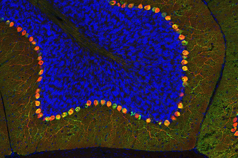
Below, Ellen DeGennaro, working in the laboratory of Christopher Walsh, MD, PhD, created this colorful image of a thin section of tissue from an adult mouse cerebellum. The image highlights blue nuclei, and Purkinje cells in red and green. The Walsh lab studies the genetics and molecular biology underlying human neurological diseases.
To view all the spectacular images visit the Beauty of the Brain
Related Posts :
-

Promising advances in fetal therapy for vein of Galen malformation
In 2024, Megan Ingram* of California and her husband were preparing for the birth of their third child when a 34-week ...
-
The hidden burden of solitude: How social withdrawal influences the adolescent brain
Adolescence is a period of social reorientation: a shift from a world centered on parents and family to one shaped ...
-
The journey to a treatment for hereditary spastic paraplegia
In 2016, Darius Ebrahimi-Fakhari, MD, PhD, then a neurology fellow at Boston Children’s Hospital, met two little girls with spasticity ...
-
A toast to BRD4: How acidity changes the immune response
It started with wine. Or more precisely, a conversation about it. "My colleagues and I were talking about how some ...