Science Seen: New microscope reveals biological life as you’ve never seen it before
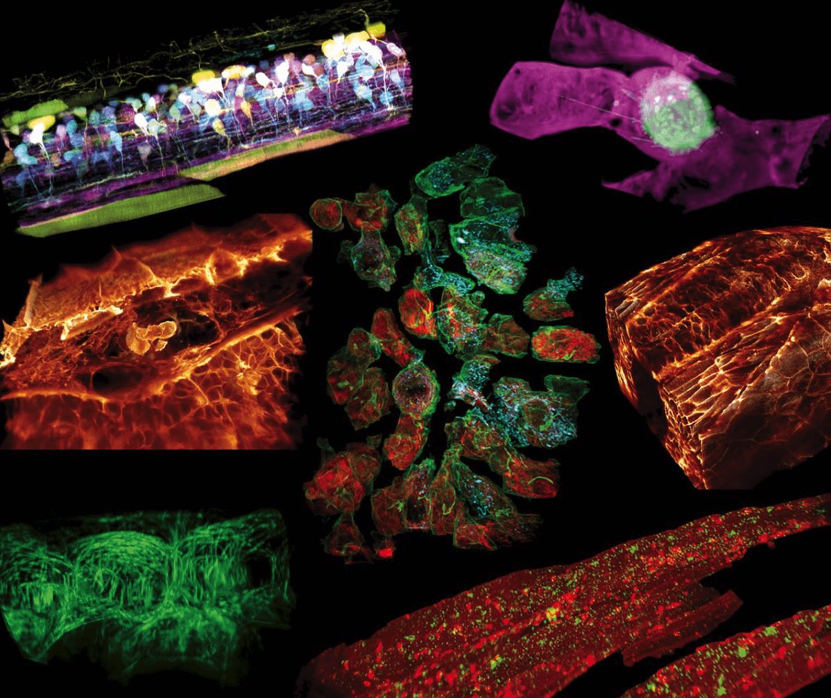
Astronomers developed a “guide star” adaptive optics technique to obtain the most crystal-clear and precise telescopic images of distant galaxies, stars and planets. Now a team of scientists, led by Nobel laureate Eric Betzig, PhD, are borrowing the very same trick. They’ve combined it with lattice light-sheet to create a new microscope that’s able to capture real-time, incredibly detailed and accurate images, along with three-dimensional videos of biology on the cellular and sub-cellular level.
The work — a collaboration between researchers at Howard Hughes Medical Institute, Boston Children’s Hospital and Harvard Medical School — is detailed in a new paper just published in Science.
“For the first time, we are seeing life itself at all levels inside whole, living organisms,” said Tom Kirchhausen, PhD, co-author on the new study, who is a senior investigator in the Program in Cellular and Molecular Medicine at Boston Children’s Hospital and a professor of cell biology and pediatrics at Harvard Medical School (HMS).
“Every time we’ve done an experiment with this microscope, we’ve observed something novel — and generated new ideas and hypotheses to test,” Kirchhausen said in a news story by HMS. “It can be used to study almost any problem in a biological system or organism I can think of.”
Seeing is believing: Here’s what the new microscope can do
So far, the team has been taking a close look at developing zebrafish because the fish have translucent skin, making it easy to capture microscopic images of their organs and tissues in vivo.
Co-first author on the study, Gokul Upadhyayula, PhD, an instructor in pediatrics at Boston Children’s and HMS, explained that zebrafish can be genetically modified so that human disease models can be studied. Already, the team has used the microscope to watch metastatic human breast cancer cells move around inside a zebrafish, revealing new insights into how cancer spreads through living tissues.


VIDEO: Kirchhausen and Upadhyayula describe their work and show some of the amazing images and 3D videos they’ve been able to capture with the microscope. Credit: Richard Groleau/Kevin Jiang/Harvard Medical School.
According to an HHMI press release, Betzig believes that adaptive optics is one of the most important areas in microscopy research today, and the lattice-light-sheet microscope, which excels at 3-D live imaging, is the perfect platform to showcase its power.
Without adaptive optics, sub-cellular microscopy is “just too damn fuzzy.”
Additional authors on the paper are researchers from Stony Brook University, University of California, Berkeley, California Institute of Technology and the University of Exeter.
This work was supported by the Howard Hughes Medical Institute, the National Institutes of Health (R01GM075252, R01DC015478, 5R00CA154870-05, 1R01GM121597-01, R01CA196884 and R35GM118149), the National Science Foundation (IOS1452928), the Carol M. Baldwin Breast Cancer Research Fund, the Damon Runyon Cancer Research Foundation, Pew Charitable Trusts, Biogen, Ionis Pharmaceuticals and a Human Frontier Science Program fellowship.
Read a related story from HMS News.
Related Posts :
-

Hope in a new home: A family’s journey with HHT
When Yeiden Pérez Camacho imagines the future, he sees himself on a basketball court. At 13, he’s already an ...
-
The hidden burden of solitude: How social withdrawal influences the adolescent brain
Adolescence is a period of social reorientation: a shift from a world centered on parents and family to one shaped ...
-
A new tool could exponentially expand our understanding of bacteria
How do bacteria — harmless ones living in our bodies, or those that cause disease — organize their activities? A new study, ...
-
AI-enabled medical devices are burgeoning, but many haven’t been tested in children
Medical devices that incorporate artificial intelligence and machine learning are proliferating. In 2013, the FDA approved fewer than 10 such devices; by 2023, ...