Exploring brain operations: Making decisions, snapping to attention, and forming memories
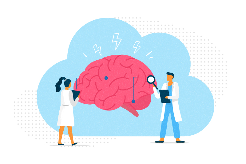
How do our brains snap to attention and orient us to the outside world — like when we’re sound asleep and the smoke alarm goes off? And when different choices confront us, how does our brain make decisions?
Two groups of researchers at Boston Children’s explored these all-important brain operations.
The first study, published February 21 in Nature, explored how the brain is organized to guide our choices.
“We wanted to understand how neurons are connected to one another in brain areas important for decision-making,” says Wei-Chung Allen Lee, PhD, a researcher with the F.M. Kirby Neurobiology Center at Boston Children’s. Lee co-led the study with Christopher Harvey, PhD, at Harvard Medical School and Stefano Panzeri, PhD, at University Medical Center Hamburg-Eppendorf.
The researchers focused on the posterior parietal cortex, an “integrative hub” that processes information gathered by multiple senses. Studying mice, they recorded brain activity as the animals chose which path to take in a maze to get to a reward (after receiving a clue). Using powerful microscopes, Lee’s lab mapped the network of connections between the neurons involved in navigating to the reward.
“We’re looking for rules of connectivity — simple principles that provide a foundation for the brain’s computations as it makes decisions,” Lee says.
When a mouse chose to turn right, a specific set of excitatory “right turn” neurons fired, they found. These neurons activated a set of inhibitory neurons that curbed activity in “left-turn” neurons — solidifying the choice to turn right. When a mouse decided to turn left, the opposite scenario played out. The researchers now hope to confirm these findings in the human brain.
Rousing an ‘offline’ brain and forming memories
The second study, led by Jordan Farrell, PhD, focused on how brains that have been zoning out wake up and focus when a situation demands it. Findings were published in Nature on March 13.
“We have found a brain mechanism for breaking up periods of mind wandering and realigning the ‘cognitive map’ back to reality,” says Farrell, an investigator in the F.M. Kirby Neurobiology Center and Rosamund Stone Zander Translational Neuroscience Center.
During sleep or periods of daydreaming, our brains replay past events in a form of synchronized activity called the “sharp-wave ripple.” This enables us to consolidate our memories.
Analyzing data from a mouse model, Farrell and colleagues from the laboratory of Ivan Soltesz, PhD, at Stanford University began exploring another, little-known neuronal activity pattern: synchronized spikes of firing in the dentate gyrus, part of the hippocampus. These spikes, they found, occur when an “offline” brain is aroused. They may help us quickly process new information and orient ourselves to what’s happening in our environment — like when the teacher suddenly called on us while we were daydreaming in class.
But dentate spikes also seem to promote associative memory — in which a sensory stimulus (say, a series of piercing beeps) is stored as a memory, so that we come to associate the noise with a smoke alarm going off and the possible need to evacuate.
Sharp-wave ripples and dentate spikes may have complementary roles, Farrell says. “The brain is toggling through these two states.”
A window on neurologic disorders?
This new knowledge about dentate spikes could open windows into some neuropsychiatric disorders. Perhaps dentate spikes affect attention and arousal in people with ADHD or post-traumatic stress. Or maybe they are altered in Alzheimer’s, disrupting formation of new memories.
Farrell is most interested in epilepsy, which is marked by synchronized spikes of excessive neuron firing. He plans to investigate the role of dentate spikes by better understanding their basic mechanisms and then manipulating the neural networks that control dentate spikes in the brains of epileptic mice. He also hopes to extend the study to children with epilepsy, in collaboration with clinicians at Boston Children’s.
“In people with epilepsy, the synchronous activity during dentate spikes could tip the brain into a pathological state,” Farrell speculates. “The dentate spikes add an extra push to the system.”
Learn more about research and innovation in Neurology
Related Posts :
-

Timing is everything: How circadian rhythms influence our brains
Why are we mentally sharper at certain times of day? A study led by Jonathan Lipton MD, PhD, at Boston ...
-
When diagnosis is just the first step: The Brain Gene Registry
Through advances in genetic sequencing, many children with rare, unidentified neurodevelopmental disorders are finally having their mysteries solved. But are ...
-
New leads for spinal cord injury: Mapping spinal-projecting neurons in the brain
Only a fraction of people who sustain a spinal cord injury fully regain their motor function. While rehabilitation can help, ...
-
Unraveling the secret to attention, one brain cell at a time: Brielle Ferguson, PhD
In college, Dr. Brielle Ferguson was initially drawn to psychology. Witnessing the impact of schizophrenia on a family member, she ...