MRI could reduce the mystery of brachial plexus injuries in infants
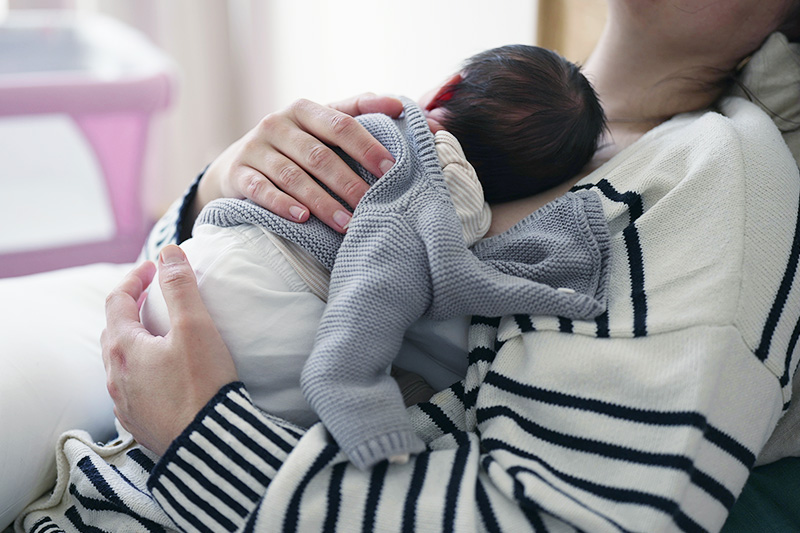
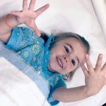
About one in 1,000 children are born with brachial plexus birth injury (BPBI), upper extremity weakness or paralysis resulting from trauma to the brachial plexus nerves during childbirth.
Most children with BPBI recover with observation and minimally invasive care, but about 30 percent have injuries severe enough to lead to long-term impairment. Thanks to recent advances in nerve repair, ruptured or avulsed nerves can often be repaired surgically. The challenge is knowing which infants would benefit.
Key takeaways
- Researchers modified MRI procedures and developed a scoring system to evaluate the extent of BPBI.
- These measures enabled more timely assessments of whether BPBI would lead to long-term loss of upper extremity function, thereby alleviating the burden on parents.
“Brachial plexus birth injury is a complicated diagnosis,” says Andrea Bauer, MD, director of the Brachial Plexus Program at Boston Children’s Hospital. Although MRI is used routinely to assess brachial plexus injury in adult patients, predicting the downstream effects of nerve damage in infants is not straightforward.
Thanks to cortical and peripheral nerve plasticity early in life, explains Bauer, an infant’s healthy nerves can compensate for damaged nerves. In other words, injury to a specific nerve does not necessarily correlate to impairment of the associated muscle. So even when an avulsion is visible on an infant’s MRI, that infant could regain function of the affected nerve root’s typical distribution without surgery.
For this reason, the current standard of care is monthly physical examinations to monitor upper extremity mobility for six to nine months. The delay adds to the stress of parents who are already dealing with an unexpected diagnosis. Further, many families must travel long distances for regular appointments at a brachial plexus center like the one at Boston Children’s.
Mapping BPBI through non-sedated MRI
The multi-center NAPTIME (non-anesthetized plexus technique for infant MRI evaluation) study aimed to develop a more timely way to determine whether BPBI is severe enough to warrant surgery.
Families of 102 infants receiving care at one of three brachial plexus centers — Boston Children’s, Gillette Children’s Hospital, and Shriners Hospitals for Children Northern California — participated in the study.
Infants were between four- and 16-weeks old — an age at which it’s too soon to know if upper extremity function will return spontaneously or with therapy. Each infant underwent a non-sedated, non-contrast MRI. The researchers then continued to provide care based on standard protocols.
Innovations in BPBI diagnosis
As part of the study, the NAPTIME research team made some important modifications to overcome known barriers to using MRI to assess brachial plexus injury in infants:
Non-sedated MRI
One of the barriers to MRI in young children is that patients must lie still during scanning. Anesthesia can prevent squirming but carries significant risks in young patients.
By narrowing down the number of MRI sequences necessary to detect BPBI, researchers were able to gather the information they needed in six minutes. They also employed a “feed and wrap” technique, in which a parent fed and swaddled their infant immediately before an MRI was performed. This induced a relaxed state, particularly in younger subjects, making it possible to perform the six-minute MRI scan without anesthesia.
BPBI scoring
The team also developed a BPBI scoring system that measured both the number of injured nerves and the severity of those injuries. Healthy nerves may compensate for damaged nerves, but this inherent ability reaches its limit when multiple nerves are severely damaged. As expected, higher scores correlated to a higher likelihood that arm function would not return without surgery. These results were published in the Journal of Bone and Joint Surgery.
A new tool in the BPBI toolkit
Based on the study results, the team at Boston Children’s now offers non-sedated MRI to families of infants with BPBI. “Most families want information about what’s going on inside, if we can gather it without harming their baby,” says Bauer. She hopes other centers will feel encouraged to adopt the practice based on the study results that demonstrate the feasibility of providing answers sooner.
In the longer term, the MRI images gathered through the study may also help shed light on shoulder function in children with BPBI. By looking at shoulder outcomes in older study participants and comparing them to patterns of injury detected in the MRIs, researchers are hoping to find whether certain patterns of injury correlate to specific longer-term outcomes. If so, brachial plexus specialists may have yet another tool to optimize their patients’ future upper extremity function.
Funding for this study came through Shriners’ Hospitals for Children.
Learn more about the Brachial Plexus Program or refer a patient
Related Posts :
-

Broken signals: Things you may not know about nerve injury
When Dr. Andrea Bauer talks about nerve injuries, she talks about phone cords. A damaged phone cord transmits staticky or ...
-
With a dose of health equity, brachial plexus study enrolls more patients
What drives a parent to say yes or no to enrolling their child in research? When a surprisingly high percent ...
-
Brachial plexus birth injury: Harper’s right arm
When Harper Jane Stalker was born with a limp, unmoving right arm in 2016, her parents had never heard of brachial ...
-
Fingers, shoulders, and everything in between: Three upper extremity surgeons and their relentless quest for solutions
It’s 6 a.m. and the surgeons in the Hand and Orthopedic Upper Extremity Program at Boston Children’s Hospital ...