A new tool could exponentially expand our understanding of bacteria
How do bacteria — harmless ones living in our bodies, or those that cause disease — organize their activities? A new study, combining powerful genomic-scale microscopy with a technical innovation, captured which genes bacteria turn on in different situations and in different spatial environments. The technology, described January 23 in Science, promises to take the study of bacteria to the next level.
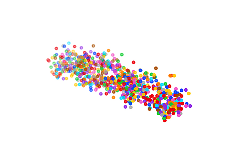
Jeffrey Moffitt, PhD, and colleagues in the Program in Cellular and Molecular Medicine (PCMM) at Boston Children’s Hospital applied MERFISH, a molecular imaging technique Moffitt helped develop, to profile messenger RNAs — representing thousands of genes — in thousands of single bacteria simultaneously. MERFISH also captured spatial information, revealing how spatial factors influence what genes bacteria turn on — something that had never been done before.
The team first had to overcome a major barrier to studying bacterial RNAs, also known as the bacterial transcriptome. Because bacterial cells are tiny, their RNAs are crammed tightly inside and mingle together, making it hard to image and identify them. “It was a complete disaster, we couldn’t see anything,” says Moffitt.
Borrowing a technique developed in the laboratory of Ed Boyden, PhD, at MIT— expansion microscopy — they embedded the samples in a special hydrogel. They anchored the RNAs to this gel, and changed the chemical buffer in the gel. This triggered it to swell, expanding the sample 50- to 1000-fold in volume. “All the bacterial RNAs become individually resolvable,” Moffitt says.
Why measure bacterial gene expression?
Until now, scientists have averaged bacterial behavior across a given bacterial population. The ability to determine what genes individual bacteria are using can give powerful new insights into bacterial interactions, virulence, stress responses, the ability to resist antibiotics, the ability to form biofilms like those in catheters, and more.
“We now have the tools to answer fascinating questions about host-microbe and microbe-microbe interactions,” Moffitt says. “We can explore how bacteria might communicate and compete for spatial niches and define the structure of microbial communities. And we can ask how pathogenic bacteria adjust their gene expression as they infect mammalian cells.”
Bacterial-MERFISH can also provide insights on bacteria that are difficult to grow in a culture dish. “Now we don’t have to culture them, we can just go image them in their native environment,” Moffitt says.
Single-cell insights
Several experiments the team performed illustrate the kinds of questions that bacterial-MERFISH can answer. For example, Moffitt and colleagues were able to show that individual E. coli, when starved of glucose, try utilizing alternative food sources one after another, altering their gene expression in a specific sequence. Taking a series of genomic snapshots over time enabled the team to piece together this survival strategy.
The team also got insights into how bacteria organize their RNAs within their cells, which may be important in how different aspects of gene expression are regulated. Finally, they showed that intestinal bacteria tap different genes depending on their physical location in the colon.
“The same bacteria could be doing very different things over a space of tens of microns,” Moffit says. “They’re seeing different environments and responding differently to them. It was very difficult to address such variation before, but now we can answer the types of questions people have been dreaming about.”
The paper’s coauthors were Ari Sarfatis, Yuanyou Wang, PhD, and Nana Twumasi-Ankrah in the Moffitt Lab.
Learn more about the Moffitt Lab and research in the Program in Cellular and Molecular Medicine (PCMM).
Related Posts :
-

A journey through the intestine during colitis, cell by cell
Inflammatory bowel disease (IBD), causing devastating abdominal pain, persistent diarrhea, and rectal bleeding, is hard to control with current treatments. ...
-
Tracking influenza in its first battleground: The nose
The answer to curbing influenza could be right under our noses — or, more accurately, inside them. New research maps happenings ...
-
Piecing together the preterm infant microbiome
The human microbiome — the collection of microbes living in the gut — is now recognized as an important contributor to health ...
-
Real-time genomic surveillance of bacteria could improve antibiotic therapy
Antibiotic-resistant bacterial infections are increasingly hard to treat, causing more than a million deaths annually around the world. Hospitalized patients ...