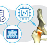
3D imaging could become standard practice in orthopedics. Here’s how.
It took a trained eye to see the abnormality on the patient’s X-ray. There, hidden behind the acetabulum was the shadow of a small bone spur.
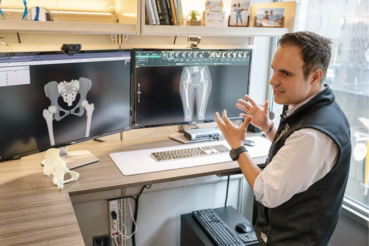
“At first glance, this looks like a normal, healthy hip,” says Young-Jo Kim, MD, PhD, director of the Child and Young Adult Hip Preservation Program at Boston Children’s Hospital. “But here you can see the tip of the bone spur at the back of the joint — which is unusual.” Kim goes on to point out that the socket is covering the femoral head more than usual, but that too is subtle, something a less experienced orthopedist could easily miss.
When he pulls up a 3D model of the same hip, the issues on the screen are far easier to spot. The bone spur that was almost imperceptible on X-ray is clearly visible, as are red areas indicating areas of bone impingement. Kim points to a green line on the femoral head. “That’s normally where the edge of the socket should be,” he says. Instead, the line is well inside the socket.
For the past several decades, 3D images have helped orthopedic surgeons visualize complex orthopedic conditions. They can also help younger surgeons more accurately identify musculoskeletal issues and patients understand the source of their pain. Time and cost, however, have been limiting factors. Typically, an imaging technician generates 3D images from MRI or CT scans. Depending on the department’s workload, this can take a week or longer.
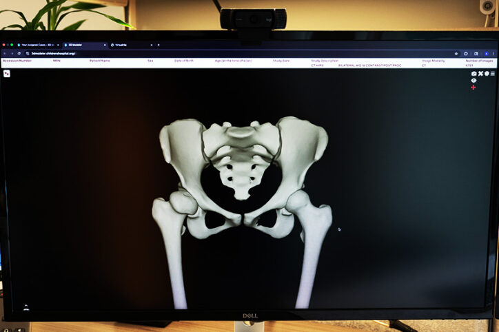
3DModelerTM, a new innovation at Boston Children’s, could change this picture.
Timely, cost-effective access to 3D imaging
3DModeler is an extension of VirtualHip, a software program developed at Boston Children’s that uses 3D imaging and artificial intelligence (AI) to support the diagnosis and treatment of hip conditions. After its successful rollout in 2023, leaders in the Orthopedics and Sports Medicine Department wanted to make 3D imaging a standard of care in every area of orthopedics and sports medicine. To do so, they’d need a whole-body 3D system.
The goal was ambitious. Ata Kiapour, PhD, director of Boston Children’s Musculoskeletal Digital Innovation & Informatics Program describes the challenges. “As a tertiary referral center, Boston Children’s treats hundreds of patients with rare and complex conditions,” he says. Not only that, but given the hospital’s pediatric population, some patients’ bones are soft and immature, while others are fully ossified.
The system would therefore need to be robust enough to model complex structural abnormalities while taking the maturity of a given patient’s bone into account. To be most useful, it would also need to produce these models quickly and at minimal cost.
The team took advantage of recent advances in AI models and infrastructure to meet each of these challenges. “We trained an AI model to create 3D geometries of bones at different stages of development,” says Kiapour. They also trained the program to accurately model patients’ soft tissues.
A clear view of cartilage
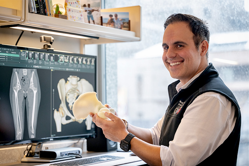
Kiapour pulls up a 3D model of a knee with osteochondritis dissecans. Using the 3DModeler, he rotates the image, pointing out bones and tissues.
“Here you can see the distribution of cartilage,” he says, indicating the base of the femur. “If there’s damage, you can see that.” And sure enough, near the joint, a small lesion is visible where a piece of cartilage has separated from the bone. While an experienced radiologist or surgeon would likely have seen the lesion on MRI, it’s far more visible, particularly to an untrained eye, on the 3D model.
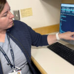
“We were able to create this model from an MRI within five minutes using 3DModeler,” says Kiapour. Once the system is up and running, clinicians will be able to generate 3D models from their computers and handheld devices — no imaging technician needed. If they want to print a 3D model, for teaching or patient-education purposes, the computer-generated models can support this.
A 3D future for orthopedics
As of now, 3DModeler can generate 3D models of any part of the body from a CT scan. If the original image is an MRI, the software can currently create 3D models of a patient’s hips, knees, or ankles. Work is underway to expand this list to include the whole body as well.
3DModeler was released for beta-testing in Boston Children’s Orthopedics and Sports Medicine Department this summer. A period of gathering clinician feedback is underway, and the team expects that within a year, 3D modeling will be a standard part of clinical practice for Boston Children’s orthopedic patients, supporting clinical decision making for physicians at all levels of expertise.
“Having a better visualization tool improves both diagnosis and surgical planning,” says Kim. “You can see very clearly what the problem is and how to fix it.”
And while AI made the technology of 3DModeler possible, having clinical leaders willing to invest in non-traditional research was just as critical.
“Thanks to the vision of our senior leaders, including Dr. Kim, Boston Children’s may be the only orthopedic department in the world to have deployed something like this in our clinics,” says Kiapour.
Learn more about the Musculoskeletal Digital Innovation & Informatics Program, Child and Young Adult Hip Preservation Program, and Orthopedics and Sports Medicine Department.
Related Posts :
-

AI could change the way we look at hip preservation
Orthopedic surgeons and biomedical engineers are trained to approach adolescent and young adult hip pain from two different perspectives. Surgeons ...
-
Ask a sports medicine specialist: Why are ACL tears so common among female athletes?
When an athlete is sprinting after an opponent who suddenly stops or changes direction, their anterior cruciate ligaments (ACLs) make ...
-
MRI could reduce the mystery of brachial plexus injuries in infants
About one in 1,000 children are born with brachial plexus birth injury (BPBI), upper extremity weakness or paralysis resulting from trauma ...
-
Engineered cartilage could turn the tide for patients with osteoarthritis
About one in seven adults live with degenerative joint disease, also known as osteoarthritis (OA). In recent years, as anterior ...