Monitoring for breast cancer after childhood chest radiation: When and how?
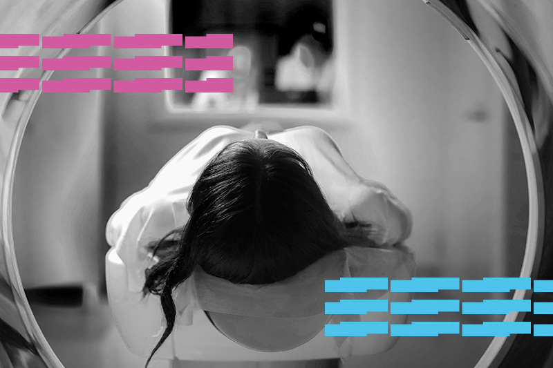
Chest radiation is used to treat children with Hodgkin and non-Hodgkin lymphoma as well as lung metastases in various solid tumors. But radiation itself is a potential cancer risk. That includes an increased risk for breast cancer later in life. Girls receiving chest radiation for childhood cancer face a breast cancer risk as high as 30 percent by age 50.
· Girls receiving chest radiation for childhood cancer face up to a 30 percent breast cancer risk by age 50.
· A simulation finds starting at age 30 with MRI and mammography to be the preferred approach to screening.
· Starting at age 25 prevented slightly more deaths, but entailed more testing and emotional stress.
What is the best strategy for catching these breast cancers early? Annual screening with mammography and breast MRI is recommended. But practices vary from location to location, and the benefits, harms, and costs are unclear.
A study led by Jennifer M. Yeh, PhD, in the Division of General Pediatrics at Boston Children’s Hospital, used modeling to compare different approaches. The findings were published yesterday in the Annals of Internal Medicine.
Comparing screening approaches
“Randomized clinical trials are often thought of as the ‘gold standard,’ but they aren’t always feasible for screening studies,” says Yeh. “That’s especially true in rare high-risk groups such as survivors of childhood cancer. However, those developing care guidelines need information on the potential benefits and harms of screening. Decision modeling can provide important insights.”

Yeh, with collaborators across the country, used two breast cancer simulation models, part of the Cancer Intervention and Surveillance Modeling Network (CISNET).
Both models were developed to guide screening recommendations for women who are not cancer survivors. To adapt them for cancer survivors, the researchers used information from the Childhood Cancer Survivor Study (CCSS). That study looked at outcomes, including breast cancer, in more than 24,000 survivors of childhood and adolescent cancers diagnosed between 1970 and 1999.
The models evaluated the following annual screening strategies:
- no screening
- digital mammography and breast MRI, starting at age 25 (as per current recommendations of the Children’s Oncology Group), 30 or 35
- MRI only, starting at age 25, 30, or 35.
The models assumed that women who were screened continued to be screened until age 74, and that those diagnosed with breast cancer received the best therapy available at the time.
All annual breast cancer screening approaches save lives
In the simulation, Yeh showed that without screening, childhood cancer survivors who had received chest radiation had a 10 to 11 percent lifetime risk of dying from breast cancer. By comparison, women in the general population had a 2.5 percent risk.
Compared with no screening, all annual screening strategies prevented more than half of breast cancer deaths, according to the models. A combined approach of both breast MRI and mammography, beginning at age 25, prevented the most deaths (an estimated 56 to 71 percent). MRI alone prevented slightly fewer (56 to 62 percent).
Factoring in quality of life
While starting at age 25 offered a slight survival benefit, it also meant more screening tests, more false-positive findings, and more breast biopsies that turned out to be benign. For example, the researchers estimate that the average survivor screened with both MRI and mammography would have four to five false-positive screens and one to two breast biopsies over the course of her lifetime.
When the emotional stress and costs of additional screening and testing were factored into the analysis, starting at age 30 was the preferred strategy; relatively little was gained by starting at age 25.
Bottom line: Either strategy reduced the risk of dying from breast cancer by at least half, with or without the addition of mammography to MRI.
Reassurance about early screening
Lisa Diller, MD, chief medical officer of the Dana-Farber/Boston Children’s Cancer and Blood Disorders Center, is an expert in the care of childhood cancer survivors and a co-author of the study.

“Breast cancer screening is one of the most important issues that oncologists should discuss with survivors of childhood chest radiation,” Diller says. “Having these data informs discussions with young women who are facing screening at a very young age. It both reassures them that waiting until age 30 might be reasonable and impresses upon them that this screening could be life-saving.”
One limitation of the study was that it used data from child cancer survivors diagnosed between 1970 and 1986. Since then, cancer treatment has changed, including decreased doses and improved delivery of radiation. And breast cancer monitoring now includes digital breast tomosynthesis, also known as 3D mammography.
“Our findings based on modeling suggest that even if breast cancer risk is cut in half with more recent changes in radiation dose and delivery, early initiation of screening still remains favorable for these high-risk survivors,” Yeh says. “Ensuring survivors are aware of and have access to screening can save lives.”
The study was funded by the American Cancer Society, the National Cancer Institute, a Cancer Center Support grant and the American Lebanese-Syrian Associated Charities.
Learn more about research at Dana-Farber/Boston Children’s.
Related Posts :
-

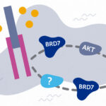
BRD7 research points to alternative insulin signaling pathway
Bromodomain-containing protein 7 (BRD7) was initially identified as a tumor suppressor, but further research has shown it has a broader role ...
-
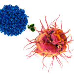
Exposing a tumor’s antigens to enhance immunotherapy
Successful immunotherapy for cancer involves activating a person’s own T cells to attack the tumor. But some tumors have ...
-
Combining CAR-T cells and inhibitor drugs for high-risk neuroblastoma
Chimeric antigen receptor (CAR)-T cell therapy is a potent emerging weapon against cancer, altering patients’ T cells so they ...
-
When diagnosis is just the first step: The Brain Gene Registry
Through advances in genetic sequencing, many children with rare, unidentified neurodevelopmental disorders are finally having their mysteries solved. But are ...